The modern MRI system is an amazing blend of diverse technologies, based on the realization that a small but critical aspect of the spin of hydrogen nuclei could be leveraged to provide a safe, non-invasive way to see inside a human body.
Part 2 of this FAQ looks at the physics and other advances that led to the MRI as we know it.
Q: What are the developments which lead to the MRI?
A: Obviously, no single person or team invented the MRI in isolation; it is the culmination of decades of scientific progress and understanding. MRI technology begins with the discovery of a quantum-physics phenomenon called nuclear magnetic resonance (NMR) in 1937 by Isidor I. Rabi, a Polish-born American physicist.
Q: What was the scope of his discovery?
A: He showed that atomic nuclei indicate their presence by absorbing or emitting radio waves when exposed to a sufficiently strong magnetic field. For this work, he received the Nobel Prize in Physics in 1944.
QL: What was the next step?
A: By 1946, Felix Bloch at Stanford University and Edward Purcell at Harvard University independently took the NMR phenomenon and extended it beyond nuclei to properties of atoms and molecules in solids and liquids. Instead of individual atoms or molecules of Rabi’s molecular beam, this became the basis for NMR-based spectroscopy to study the composition of chemical compounds (they received a Nobel Prize in Physics for this work in 1952).
Q: All this still seems far away from the MRI — what happened next?
A: Subsequently, Paul Lauterbur, a professor of Chemistry at the State University of New York at Stony Brook, proposed a technique and experiments which took the single dimension of NMR-based spectroscopy to two-dimensional imaging. In 1973 his work produced the first NMR image (of a test tube). Around the same time, Peter Mansfield of Nottingham, England, showed how gradients in the magnetic field could be mathematically analyzed, making a useful, fast-imaging technique possible. The two received the 2003 Nobel Prize in Physiology or Medicine for their work.
Q: It feels like we are getting closer to the actual MRI system–is this the case?
A: The concept and realization of a complete resonance-based imaging system were developed and implemented by Dr. Raymond Damadian, a physician and experimenter working at Downstate Medical Center in Brooklyn, New York. He realized that because tumors, for example, contain more water and thus more hydrogen atoms than healthy tissue, there might be a way to use an atomic-level response to see the difference and create images.
Q: Did he stumble on this by accident?
A: Not at all. Damadian found that different kinds of animal tissue emitted signals that vary in length, and that the signals from cancerous cells last much longer than those from non-cancerous tissue. He filed for and received a patent in 1974 for an “Apparatus and Method for Detecting Cancer in Tissue,” the first MRI-related patent.
Q: Did his work stop with the patent?
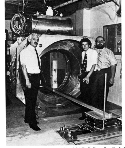
A: No, he and his team completed construction of a whole-body MRI scanner in 1977. They built the entire machine themselves, including winding the magnet coils and plumbing the superconducting-magnet subsystem, Figure 1. The first test was a failure, but Damadian and his team realized the problem: he was physically too large for the sensor array.

Q: So what did they do?
A: One of his graduate students, Dr. Larry Minkoff, volunteered to take his place, and after nearly five hours, the first human scan was complete (Figure 2; Figure 3 is a scan of the same area (approximately)with a modern MRI system, while Figure 4 is the same area from a standard anatomy drawing. Reference 1 is Dr. Damadian’s overview paper on the construction of the machine, the underlying analysis, and the data.


Q: What about commercialization?
A: In 1978, Damadian formed FONAR (“Field Focused Nuclear Magnetic Resonance”), and in 1980, he produced the first commercial unit. However, for technical reasons, he had to abandon his original MRI technique in favor of a variation developed by Lauterbur and Mansfield.
Q: Sounds like a straightforward patent and IP situation, but was it?
A: Not at all; this is where things became nasty. Damadian and Fonar obtained royalties on their patents and settled with many large companies to use the technology. General Electric would not acknowledge the validity, however, but Fonar eventually prevailed (it took over a decade of litigation) and received a $129 million ruling against GE.
Q: End of story, right?
A: Not at all! The Nobel Prize awarded to Lauterbur and Mansfield did not include Damadian (note that Nobel rules allow for up to three recipients). The dispute over “proper credit” for MRI has gone on for many years, with vocal adherents on both sides. The American Physical Society (APS) page on MRI (Reference 2) gives full names and details for the fundamental research but suddenly changes to an anonymous, passive tone when it gets to specifics of the first MRI. It makes no mention of Damadian; similarly, a detailed and lengthy reference from The Journal of Cardiovascular Magnetic Resonance mentions him once, and then only as part of a list of contributors to MRI development.
Q: Was all this controversy “behind the scenes”?
A: To the contrary: adherents on both sides of the controversy mounted public campaigns, with letters in scientific journals, press conferences, debates at conferences and even full-page newspaper ads (this was before the Internet) — all of which was a rather unusual spectacle for the scientific community.
Q: What’s the status of MRI systems now?
A: Advances in super magnets, computing power, algorithms, video display resolution, RF generation and control, and many other technical areas have advanced MRI to new approaches and capabilities. For example, since protons in different body tissues relax and return to their normal spins at different rates when the transmitted RF pulse is switched off, the scanner can be adjusted to distinguish among tissues. Additional magnetic fields can be used to localize body structures in three dimensions, and multiple radio-frequency pulses can be transmitted in sequence to highlight specific tissues or abnormalities.
Q: What are some other MRI enhancements?
A: Many application-specific MRI variations now exist, such as diffusion MRI and functional MRI. Diffusion MRI measures how water molecules diffuse through body tissues; some diseases, such as a stroke or tumor, can restrict this diffusion, so this method is often used to diagnose them. Functional MRI (fMRI) measures changes in blood flow in different parts of the brain by assessing blood-oxygen-level-dependent (BOLD) contrast. As neurons use more oxygen when they’re active, these BOLD signals are an indication of brain activity, and fMRI has had a major impact on studies of the brain.
Other advances include mobile machines which can be carried in a truck and set up where needed and even “open” designs where the subject is not surrounded by the RF coils (which can be somewhat claustrophobic and unsettling. Scan speeds have also been reduced by orders of magnitude, so the subject does not have to lie still for minutes or even hours.
Q: Is there a larger lesson in the development of MRI, from basic atomic physics to the huge instruments in widespread use?
A: MRI is an excellent example of where developments across multiple disciplines (atomic-level insight, supercooling and intense magnetic fields, digital algorithm processing, low-noise RF amplifiers, power RF amplifiers, to cite just a few) enable unforeseen advances in unrelated fields (medical imaging). The technology also serves up an example of how a fundamental discovery with no apparent practical use can become the basis for a revolutionary new technique, product, and industry.
References
- Damadian, “Field-focusing n.m.r. (FONAR) and the formation of chemical images in man”
- APS News, “MRI Uses Fundamental Physics for Clinical Diagnosis”
- Two Views, “The History of MRI”
- Thought Co., “Magnetic Resonance Imaging MRI”
- US National Library of Medicine, National Institutes of Health, “Magnetic resonance imaging”
- Fonar, Inc, “MRI History Timeline”

